Human Body Terminology
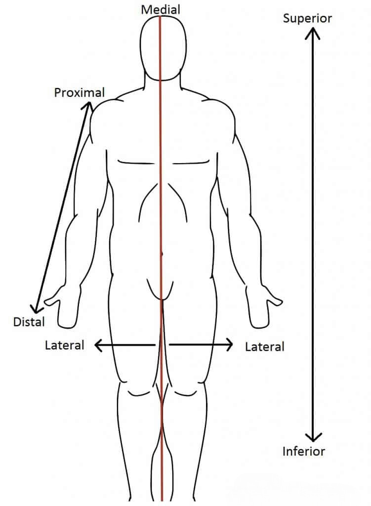
Standard Anatomical Position: Standing upright with the head and eyes looking straight ahead, arms resting at the sides, palms facing outward, thumbs pointing away from the body, feet flat on the ground, and toes directed forward.
- Anterior: Situated at the front or towards the front.
- Posterior: Positioned at the back or behind.
- Distal: Located farther away from the point of origin or attachment.
- Proximal: Positioned nearer to the point of origin or attachment.
- Superior: Situated above or higher in position.
- Inferior: Located below or lower in position.
- Lateral: Directed toward the side, farther from the body’s midline.
- Medial: Oriented toward the midline or center of the body.
- Ventral: Directed toward the underside or belly.
- Dorsal: Positioned closer to the upper surface or back.
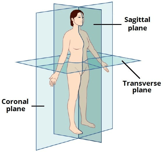
Body Planes
- Coronal Plane (Frontal Plane): Separates the body into anterior (front) and posterior (back) sections.
- Sagittal Plane (Lateral Plane): Splits the body into right and left portions.
- Midsagittal Plane: A specific sagittal plane that passes through the center of the body, dividing it into equal left and right halves.
- Transverse Plane (Axial Plane): Divides the body into upper (top) and lower (bottom) parts.
Medical Terminology
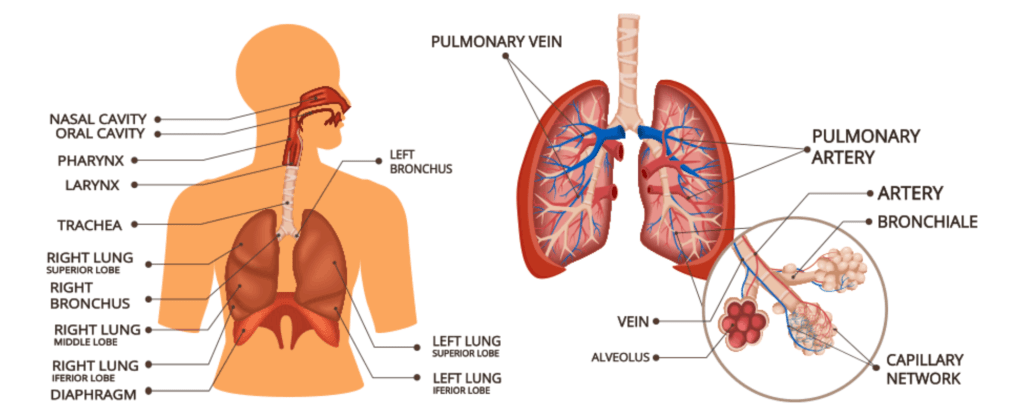
The Respiratory System
Continuous Gas Exchange: The body constantly interacts with the environment to maintain oxygen levels and expel carbon dioxide.
Breathing: The process of inhaling to draw oxygen into the lungs and exhaling to remove carbon dioxide. Inhalation introduces O₂, while exhalation eliminates CO₂.
Ventilation: The physical process of oxygen and carbon dioxide exchange within the lungs, driven by the diaphragm, intercostal muscles, and the creation of negative pressure.
Tidal Volume: The volume of air moved in and out of the lungs during a regular, unforced breath.
Residual Volume: The volume of air that remains in the lungs after a full exhalation.
Pleura: A double-layered protective membrane encasing the lungs. The visceral pleura lines the lungs’ surface, while the parietal pleura covers the inner chest wall.
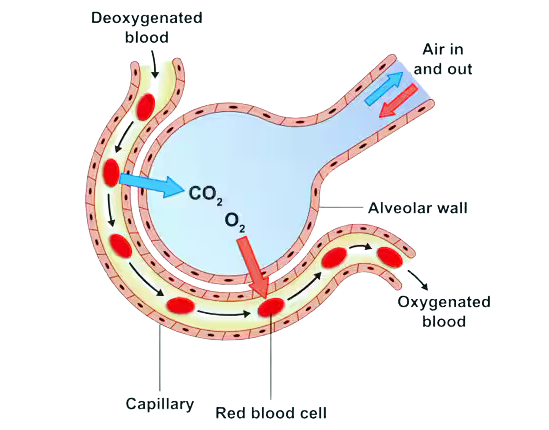
Alveolar Gas Exchange: Oxygen and carbon dioxide are exchanged between the air in the alveoli and the capillaries surrounding them.
Respiratory Control: The medulla oblongata, located in the brain, regulates breathing by tracking carbon dioxide levels and blood pH. When blood pH decreases, the respiratory rate increases to restore balance.
Alveoli: Tiny air sacs with an extensive surface area designed for efficient gas exchange via diffusion (movement from higher to lower concentrations). Type I alveolar cells within the alveoli produce surfactant, a substance that reduces surface tension to prevent alveolar collapse.
Pathologies
- Chronic Obstructive Pulmonary Disease (COPD): A collection of lung disorders that obstruct airflow and cause breathing difficulties.
- Emphysema: Damage and destruction of alveoli, impairing gas exchange.
- Chronic Bronchitis: Persistent inflammation of the bronchial tubes.
- Asthma: A condition where the airways become inflamed, constricted, and swollen due to an immune reaction to specific triggers.
- Respiratory Tract Infections:
- Upper Respiratory Infections: Impact the nose and throat, reducing air intake.
- Lower Respiratory Infections: Target the lungs and nearby pulmonary structures.
- Viral Infections: Examples include influenza, COVID-19, and the common cold.
- Bacterial Infections: Examples include tuberculosis and pertussis (whooping cough).
- Pneumonia: A bacterial or viral infection affecting the alveoli, often seen in individuals with compromised immune systems.
The Cardiovascular System
- Primary Function: To distribute blood and essential substances such as nutrients, hormones, oxygen, and gases, while removing waste products throughout the body.
Circulatory System Loops
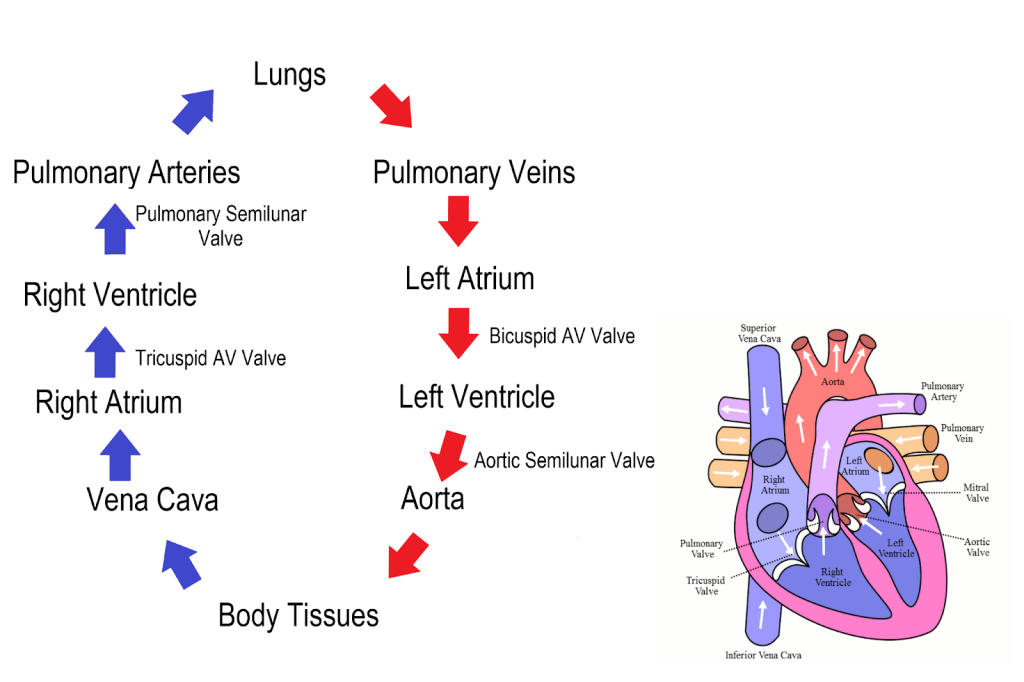
- Pulmonary Loop: Transports deoxygenated blood from the heart to the lungs, where it expels carbon dioxide (CO₂) and absorbs oxygen (O₂). The oxygen-rich blood then returns to the heart.
- Systemic Loop: Delivers oxygenated blood from the heart to the entire body and carries deoxygenated blood back to the heart.
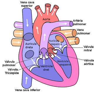
Heart Contraction Phases
- Systole: The contraction phase of the heart muscles.
- Diastole: The relaxation phase of the heart muscles.
- Heart Sounds:
- “Lub” Sound: Caused by the closing of the mitral and tricuspid valves.
- “Dub” Sound: Created when the semilunar valves close, preventing blood from flowing back into the ventricles.
Heart Regulation
- Sinoatrial Node (SA Node): Known as the heart’s “pacemaker,” it regulates heartbeats via electrical impulses.
Blood Vessels
- Arteries: Carry oxygen-rich blood away from the heart. They have thick, elastic walls to withstand high pressure.
- Veins: Transport deoxygenated blood back to the heart. Their walls are thinner than arteries.
- Capillaries: The smallest blood vessels, connecting arterioles (small arteries) to venules (small veins) and facilitating the exchange of oxygen, nutrients, and waste.
- Arterioles: Small arteries that branch into capillaries.
Pathologies
- Heart Attack: A condition where blood flow to the heart is obstructed or halted.
- Heart Failure: When the heart is unable to pump blood or fill with blood effectively.
- Arrhythmia: An abnormal or irregular heart rhythm.
- Atherosclerosis: The accumulation of plaque and white blood cells in the arteries, restricting blood flow.
- Hypertension: Elevated blood pressure levels.
- Hypotension: Abnormally low blood pressure.
- Stroke: A disruption in the blood supply to the brain.
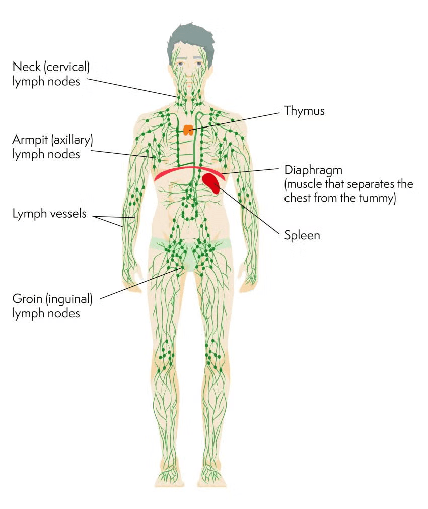
The Lymphatic System
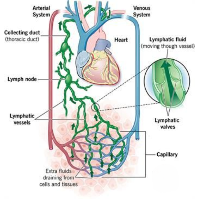
- A part of the body’s open circulatory system that works in conjunction with the cardiovascular system.
- Helps transport substances between blood and cells by clearing interstitial fluid (the fluid surrounding cells).
- Plays a vital role in the immune system by circulating white blood cells.
- Lymph: Also called lymphatic fluid, it is the surplus fluid drained from tissues and cells. It transports lymphocytes (white blood cells).
- Lymph nodes: Small, bean-shaped structures that filter and purify lymph while producing and storing lymphocytes.
- Lymphatic vessels: A network of capillaries and tubes that carry lymph throughout the body.
- Collecting ducts: Structures where lymphatic vessels deposit lymph, including the right lymphatic duct and the left lymphatic (thoracic) duct, which return the fluid to the bloodstream.
- Spleen: The body’s largest lymphatic organ.
- Tonsils: Lymphatic tissues that capture pathogens from ingested food and inhaled air.
- Peyer’s patches: Clusters of lymphatic tissue in the small intestine lining that combat intestinal bacteria.
- Appendix: Contains lymphatic tissue to help neutralize bacteria before it penetrates the intestinal wall.
Pathologies
- Lymphedema (Edema): Swelling caused by fluid buildup due to a blockage in the lymphatic system, often resulting from scar tissue or fluid accumulation.
- Lymphadenopathy: Swollen or enlarged lymph nodes, typically caused by infections, inflammation, or cancer.
The Nervous System

- Neuron: The basic functional unit of the nervous system, responsible for transmitting and receiving electrical signals.
- Axon: A long extension of the neuron that transmits information across distances.
- Dendrites: Branch-like structures extending from the neuron that collect signals from nearby cells.
- Synapse: A small gap where neurons communicate with one another without direct contact.
- Neurotransmitters: Chemical messengers that enable communication across synaptic gaps.
- Axon terminal: The neuron’s end points, shaped like small buttons, where synapses are formed.
- Glial cells: Supportive cells that protect, nourish, and provide structural support to neurons.
- Schwann cell: A type of glial cell that produces myelin, a fatty layer that insulates neurons and accelerates electrical signal transmission.
- Nodes of Ranvier: Gaps in the myelin sheath that help speed up electrical impulses.
Two main divisions of the nervous system:
Central Nervous System (CNS)
Includes the brain and spinal cord.
- Governs cognition, emotions, and behaviors.
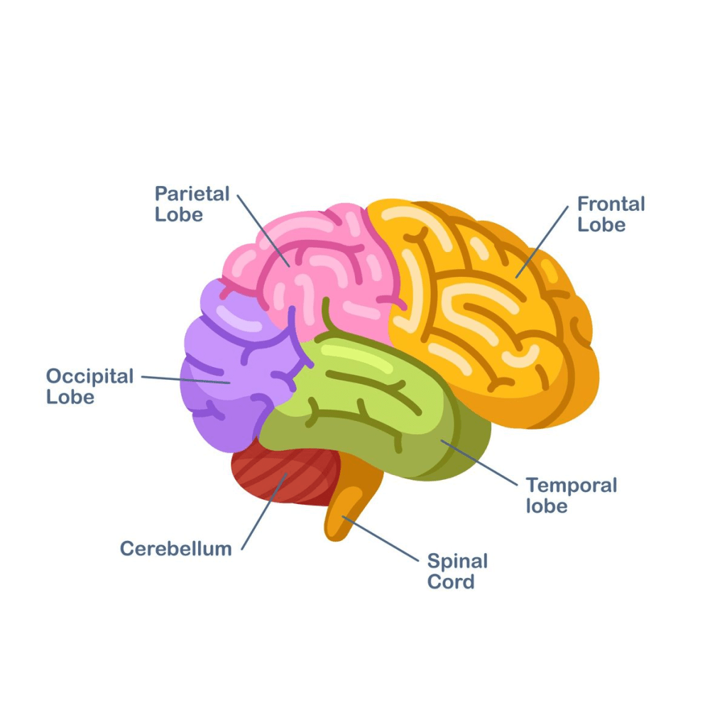
The brain is divided into specific regions called lobes:
- Frontal lobe: Controls voluntary movement, memory, self-regulation, and expressive language.
- Occipital lobe: Processes visual input.
- Temporal lobe: Handles language comprehension and emotional responses.
- Parietal lobes: Interprets sensory information, including touch, smell, and pain.
- Cerebral cortex: The brain’s outermost layer.
- Sulci: Grooves or “valleys” on the brain surface.
- Gyri: Raised areas or “ridges” between sulci.
- White matter: Composed of myelinated nerve fibers.
- Grey matter: Made up of unmyelinated nerve fibers.
Peripheral Nervous System (PNS)
Comprises all nerves outside the brain and spinal cord.
- Acts as a communication bridge between the CNS and the rest of the body.
Functional classifications:
- Afferent (sensory) neurons: Relay sensory data, such as smell, taste, touch, and pain, to the CNS.
- Efferent (motor) neurons: Transmit signals from the CNS to muscles.
- Autonomic nervous system (ANS): Oversees involuntary functions like digestion, breathing, and heart rate. It regulates the “fight or flight” response via adrenal glands.
- Somatic nervous system (SNS): Controls voluntary actions such as walking, running, and throwing.
Key features and processes:
- Brain signals travel down the spinal cord before engaging motor neurons at neuromuscular junctions.
- Reflex: An automatic, rapid response to external stimuli.
- Reflex arc: A direct pathway linking sensory neurons to motor neurons.
The Digestive System
Digestion: The process of breaking down food, starting as soon as food enters the mouth.
- Mechanical digestion: The physical breakdown of food into smaller pieces.
- Saliva: Moistens food and contains enzymes for chemical digestion.
- Chemical digestion: Involves enzymes breaking food into simpler chemical compounds.
- Mastication: The act of chewing.
- Deglutition: The process of swallowing.
- Bolus: A soft, chewed mass of food that travels down the esophagus.
- Epiglottis: A flap that closes off the trachea in the pharynx, preventing food from entering the airway.
- Peristalsis: Rhythmic muscle contractions that push partially digested food toward the stomach.
- Gastric sphincter: The valve controlling entry to the stomach.
Three main stomach secretions:
- Mucus: Protects the stomach lining.
- Hydrochloric acid: Creates an acidic environment for digestion.
- Pepsinogen: Converts into pepsin to break down proteins chemically.
Chyme: The semi-liquid mixture of digested food and stomach secretions that moves into the small intestine.
Small intestine sections:
- Duodenum: The first section, where bicarbonate neutralizes stomach acid, and nutrient absorption begins.
- Jejunum: The middle section, primarily responsible for absorbing folic acid.
- Ileum: The final section, where digested nutrients and fats are absorbed into the bloodstream.
From the ileum, the digested material enters the cecum and moves into the large intestine.
Large intestine: Absorbs leftover water, salts, and vitamin K.
Rectum: Stores waste, which is then eliminated through the anus.

Hormones
- Ghrelin: Triggers feelings of hunger.
- Leptin: Produces the sensation of fullness.
- Gastrin: Promotes digestion by stimulating the release of hydrochloric acid.
- Insulin: Facilitates glucose uptake into cells.
- Glucagon: Initiates the breakdown of stored glycogen into glucose.
Enzymes
- Salivary amylase: Begins starch digestion in the mouth.
- Pepsin: Breaks proteins down into larger peptide fragments.
- Amylase: Continues starch digestion in the small intestine.
- Lipase: Responsible for breaking down fats.
- Maltase, sucrase, and lactase: Convert disaccharides into monosaccharides in the small intestine.
- Peptidase: Breaks dipeptides into individual amino acids in the small intestine.
The Skeletal System
Made up of bones and tissues that support organ function, enable movement, and are involved in the production of blood cells, immune cells, calcium, phosphate, and lipids.
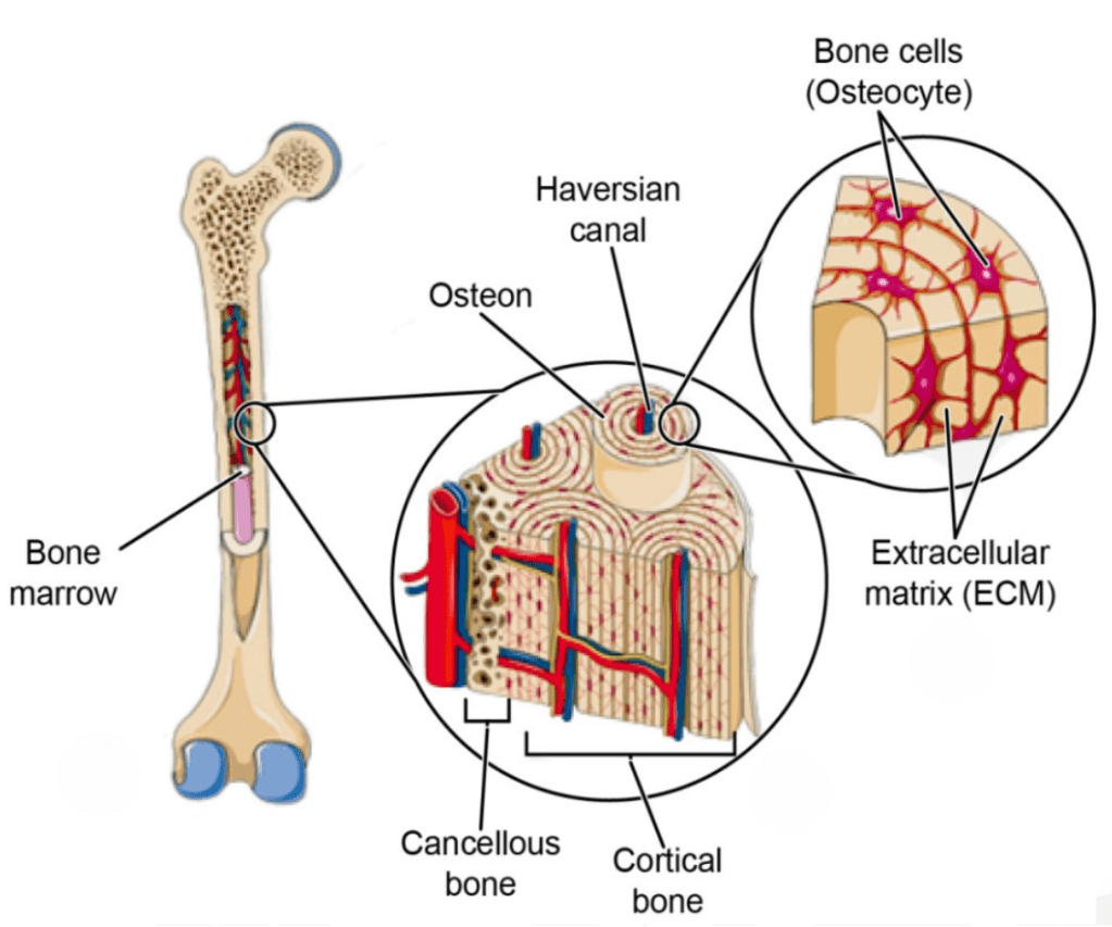
Bone structure and components:
- Outer layer: A collagen and mineral matrix that provides strength and rigidity.
- Matrix: Comprised of osteons, the functional units of bone.
- Lamellae: Compact bone layers within osteons.
- Haversian canal: Central canal surrounded by lamellae, containing blood vessels, nerves, and veins.
- Periosteum: The membrane covering the outer surface of bones.
- Volkmann’s canals: Channels that connect Haversian canals to the periosteum.
- Osteoblasts: Cells responsible for forming new bone tissue.
- Osteocytes: Mature osteoblasts embedded in the hardened bone matrix.
- Lacunae: Small cavities housing osteocytes within bone tissue.
- Canaliculi: Tiny channels connecting lacunae, facilitating communication and nutrient transfer.
- Osteoclasts: Cells on the bone surface that break down bone tissue to regulate calcium levels.
- Lining cells: Flattened osteoblasts that protect bone and help maintain calcium balance.
- Trabeculae: Spongy bone layer containing bone marrow, where red blood cell production (hematopoiesis) occurs. Bone marrow also produces lymphocytes for immune function.
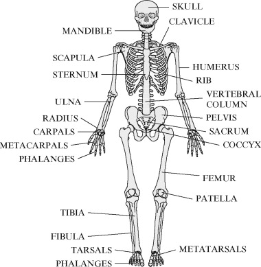
Four categories of bones:
- Long bones: Longer than wide (e.g., humerus, femur).
- Short bones: Wider than long (e.g., carpal, tarsal bones).
- Flat bones: Broad and flat, providing protection (e.g., skull, pelvis, rib cage).
- Irregular bones: Unique shapes that don’t fit other categories (e.g., vertebrae, jaw bones).
Joints and connective tissue:
Bones are connected at joints by ligaments.
- Fibrous joints: Connected by dense, collagen-rich fibers.
- Cartilaginous joints: Bound by hyaline cartilage.
- Synovial joints: Joined by synovial fluid, which lubricates and enables movement.
Pathologies
- Osteoporosis: A condition where bones lose tissue, becoming brittle and prone to fractures.
- Osteoarthritis: The degeneration of cartilage within joints.
- Rheumatoid arthritis: An autoimmune disorder targeting synovial membranes.
The Muscular System
The muscular system
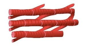
Muscle tissue comprises two primary proteins: actin (thin filaments) and myosin (thick filaments), organized in a lattice structure.
- Upon receiving a signal from a motor neuron, actin and myosin filaments slide past one another, leading to muscle contraction and shortening.
- Muscle relaxation occurs when these filaments return to their resting positions.
Adenosine Triphosphate (ATP): The energy molecule essential for both muscle contraction and relaxation.
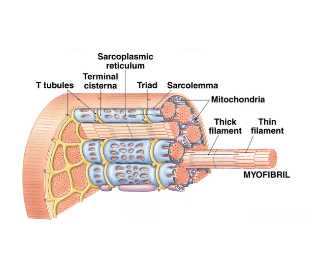
Skeletal muscle structure:
- Epimysium: Connective tissue sheath encasing the entire muscle.
- Fascicles: Bundles of muscle fibers within the muscle, each surrounded by perimysium.
- Muscle fibers (cells): Individual muscle cells encased in endomysium (a layer of areolar connective tissue).
- Sarcolemma: Plasma membrane enveloping each muscle fiber.
- Sarcoplasmic reticulum: Organelle that releases calcium ions to facilitate muscle contraction.
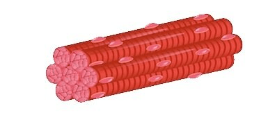
- Myofibrils: Elongated contractile threads within striated muscle cells.
- Sarcomeres: Fundamental contractile units of muscle fibers, composed of actin and myosin filaments.
Tendons: Bands of connective tissue composed of collagen fibers that attach muscles to bones.
Three types of muscle tissue
- Skeletal muscle: The only voluntary muscle type in the body, attached to the skeleton to enable movement.
- Smooth muscle: Involuntary muscle located in the esophagus, stomach, intestines, blood vessels, bladder, and bronchi. It is the weakest form of muscle tissue.
- Cardiac muscle: Involuntary muscle found exclusively in the heart, responsible for contracting and pumping blood.

Pathologies
Muscle strain: Overextension of muscle fibers, leading to discomfort, stiffness, and bruising.
Muscle sprain: Damage to ligaments surrounding a joint.
Muscular dystrophy: A genetic disorder causing gradual muscle weakening and impaired mobility.
Muscle atrophy: Reduction in muscle mass and tissue due to deterioration.
The Immune System
Serves to shield the body from harmful bacteria and viruses that lead to illnesses.
Innate Immune
General or broad in response to pathogens.
Three lines of defense:
- Skin, mucus, and secretions (acids, enzymes, salts): Act as physical and chemical barriers to block pathogens from entering the body.
- Phagocytes, specific proteins, and the inflammatory response: Target and combat pathogens that breach the initial defenses.
- Inflammatory response: Histamines are released, increasing blood flow to the affected area and boosting white blood cell (phagocyte) presence.
- Phagocytes: Engage in destroying harmful bacteria.
- Interferons: Inhibit the replication of viruses.
- Fever: Elevates body temperature to accelerate immune response.
- Natural Killer (NK) lymphocytes: Attack infected host cells harboring pathogens.
- Adaptive immune system: Tailored response that specifically targets and neutralizes particular pathogens.
Adaptive Immune
Directed at a specific pathogen.
Responses to specific pathogens:
- Cellular response: Eliminates infected cells.
- Humoral response: Targets and neutralizes pathogens in body fluids using antibodies.
- Antigen: A substance that triggers the immune response, which the body remembers if the pathogen is encountered again.
Lymphocytes:
- T-cells: Directly attack and destroy pathogens or infected cells.
- B-cells: Produce antibodies in response to antigens.
Antigen-presenting cells (APC): Digest pathogens and display their antigens to helper T-cells.
- Helper T-cells: Release cytokines and activate cytotoxic T-cells.
- Cytotoxic T-cells: Identify and destroy any cell displaying the antigen.
Memory cells: Retain information about specific antigens for future recognition.
Plasma cells: Generate antibodies to target the antigen.
Acquired Immunity
Active Immunity: Acquired through direct exposure to a pathogen or through vaccination.
- Natural: Antibodies produced after encountering a pathogen.
- Artificial: Antibodies generated through vaccination..
Passive Immunity: Gained from an external source, either naturally or artificially.
- Natural (transfer): Antibodies passed from mother to infant via breast milk.
- Artificial (transfer): Antibodies administered through immune serum treatments.
Pathologies
Autoimmune disease: A condition where the immune system mistakenly targets and damages healthy cells (e.g., lupus, psoriasis, multiple sclerosis).
Allergy: An immune reaction to substances that are typically harmless.
Human Immunodeficiency Virus (HIV): A virus that damages the body’s helper T-cells, leading to:
Acquired Immunodeficiency Syndrome (AIDS): A weakened immune system that allows infections to overwhelm the body due to insufficient immune defense.
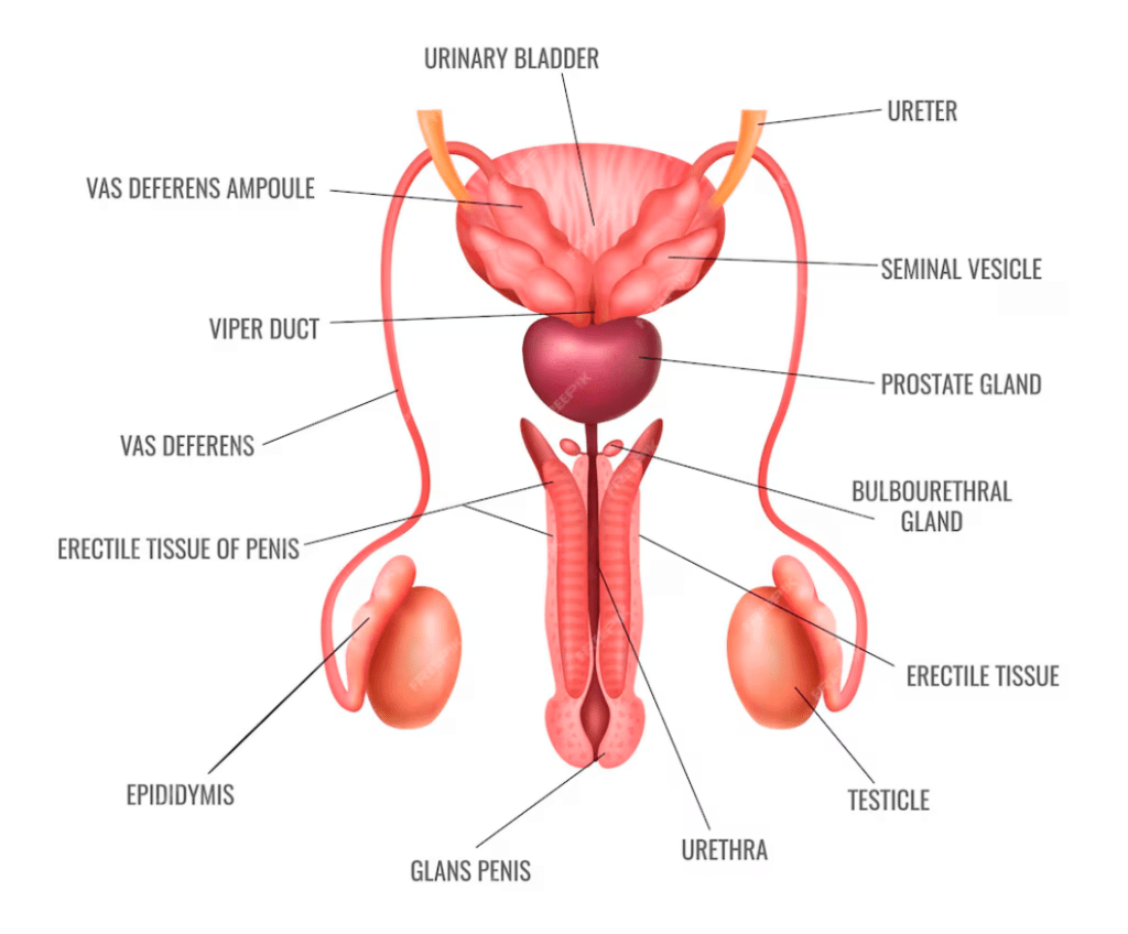
The Male Reproductive Sytem
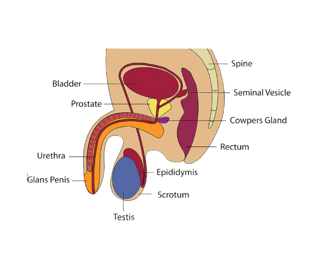
- The primary role of the male reproductive system is to generate sperm (male gametes) and deliver them to the female reproductive system.
- Sperm are created in the testes.
- The testes are located outside the body within the scrotum, a pouch-like structure.
- To support optimal sperm production, the testes need to be maintained at a temperature 2-4°C lower than the body’s normal temperature. The scrotum adjusts by contracting and relaxing, positioning the testes closer to or farther from the body.
- Epididymis: The location where sperm mature and are stored.
- Vas deferens: A narrow, elongated tube that transports sperm from the epididymis.
Three accessory glands add fluid to sperm, forming semen as it moves through the vas deferens:
- Seminal vesicles: Produce the majority of the fluid, rich in proteins, sugars, and enzymes.
- Prostate gland: Secretes an alkaline fluid that helps neutralize the vaginal tract’s acidity.
- Bulbourethral glands (Cowper’s glands): Release a lubricating, protein-rich fluid.
- Semen moves through the urethra and exits the body via the penis.
- Testosterone: The primary hormone in the male reproductive system, primarily produced by the testes and to a lesser extent by the adrenal medulla. It is responsible for the development of male reproductive organs and secondary sexual traits, such as muscle mass, facial hair, and a deeper voice.
- Spermatogenesis: The process of sperm cell formation and development.
- Erection: When erectile tissue fills with blood, causing the penis to become firm.
- Ejaculation: The forceful expulsion of semen from the male reproductive tract.
- Vasectomy: A surgical procedure in which the vas deferens are cut to induce sterilization.
- Circumcision: The surgical removal of the foreskin, often performed at birth.
Pathologies
- Prostate cancer: Cancer affecting the prostate gland, ranking as the second leading cause of cancer-related deaths in men, particularly those over the age of 65.
- Erectile dysfunction: The inability to achieve or sustain an erection, often linked to underlying health issues or psychological stress.
- Testicular torsion: A condition where the testicle twists, cutting off blood supply through the spermatic cord. This can result in testicular loss or infertility if not treated promptly.
The Female Reproductive System
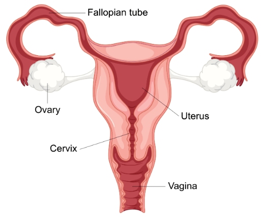
The primary function is to produce eggs (female gametes) and carry the fetus during pregnancy.
- Ovaries: the organs where eggs are produced.
- Fallopian tubes (oviducts): the site where fertilization occurs, connecting the ovaries to the uterus.
- Fertilization: the union of egg and sperm, also known as conception.
- Zygote: the initial cell formed from the fusion of egg and sperm.
- Blastocyst: a zygote that undergoes several cell divisions, forming a ball of cells ready for implantation.
- Implantation: the beginning of pregnancy when the blastocyst attaches to the uterine wall.
- Following implantation, the blastocyst becomes an embryo, and with further development, it turns into a fetus.
- Gestation: the period of pregnancy, lasting about 38–40 weeks.
- Uterus: the organ where the fetus develops during pregnancy.
- Labia majora and labia minora: outer and inner folds that lead to the vagina.
- Vagina: the birth canal and female copulatory organ, connecting the cervix to the external body.
- Endometrium: the blood-rich lining of the uterus, which sheds during menstruation if pregnancy does not occur.
- Placenta: a temporary organ formed during pregnancy that provides nutrients to the fetus and removes waste.
- Umbilical cord: connects the fetus to the placenta.
- During labor, the outer uterine layer (myometrium) contracts, pushing the fetus through the cervix and into the vagina.
- After birth, both the placenta and umbilical cord are expelled.
- Parturition: the process of giving birth.
Hormones:
- Follicle-stimulating hormone (FSH): stimulates the ovaries to produce eggs.
- Luteinizing hormone (LH): triggers the release of eggs from the ovaries during ovulation.
- Estrogen: promotes the development of female characteristics and stimulates the growth of Graafian follicles, which contain immature eggs.
Pathologies
- Endometriosis: a condition where endometrial tissue grows outside the uterus, leading to pelvic pain.
- Polycystic Ovary Syndrome (PCOS): a hormonal disorder characterized by enlarged ovaries with cysts on the edges.
- Uterine fibroids: noncancerous growths in the uterus, typically occurring during the childbearing years.
- Cervical cancer: a malignant tumor of the cervix.
- Ovarian cancer: cancer affecting the ovaries.
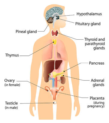
The Endocrine System
- A collection of glands that secrete hormones, which help regulate nearly every bodily function.
- Pineal gland:
- Regulates: the sleep/wake cycle (circadian rhythm).
- Produces: melatonin.
- Pituitary gland:
- Regulates: growth, blood pressure, kidney water reabsorption, body temperature, pain relief, and some reproductive processes.
- Produces: Human growth hormone (HGH), thyroid-stimulating hormone (TSH), prolactin (PRL), luteinizing hormone (LH), follicle-stimulating hormone (FSH), oxytocin, antidiuretic hormone (ADH).
- Hypothalamus:
- Regulates: pituitary activity and various metabolic functions, such as body temperature, hunger, thirst, and circadian rhythms.
- Produces: thyrotropin-releasing hormone (TRH), dopamine, growth hormone-releasing hormone (GHRH), gonadotropin-releasing hormone (GnRH), oxytocin, vasopressin.
- Thyroid gland:
- Regulates: energy usage and protein synthesis.
- Produces: thyroxine (T4), triiodothyronine (T3), calcitonin.
- Parathyroid gland:
- Regulates: calcium and phosphate levels.
- Produces: parathyroid hormone (PTH), calcitonin.
- Adrenal glands:
- Regulates: the “fight or flight” response, as well as salt and blood volume.
- Produces: epinephrine, norepinephrine, cortisol, androgens.
- Pancreas:
- Regulates: blood sugar and metabolism.
- Produces: insulin, glucagon, somatostatin.
- Testes:
- Regulates: the development of sex organs and secondary male characteristics.
- Produces: androgens (testosterone).
- Ovaries:
- Regulates: the development of sex organs and secondary female characteristics.
- Produces: progesterone, estrogen.
- Placenta:
- Regulates: pregnancy and childbirth.
- Produces: progesterone, estrogen, human chorionic gonadotropin (hCG), human placental lactogen.
Feedback mechanisms
- Positive feedback: A mechanism where hormone secretion enhances or accelerates a change, moving the system further from equilibrium.
Example: During childbirth, oxytocin is released, which intensifies cervical stretching and promotes the expulsion of the baby. - Negative feedback: A regulatory mechanism that releases hormones to restore balance and return the body to homeostasis, counteracting the changes. Most hormone regulation follows this process.
Example: The pancreas adjusts insulin secretion based on blood glucose levels.
Pathologies
- Diabetes: A condition where the body struggles to produce insulin, leading to high blood sugar levels.
- Type 1 diabetes: A chronic condition where the pancreas produces little to no insulin.
- Type 2 diabetes: A disorder that affects how the body processes sugar.
- Hypothyroidism: A condition in which the thyroid gland doesn’t produce enough thyroid hormone.
The Integumentary System
- The skin is the body’s largest organ.
- The integumentary system consists of the skin, hair, and nails.
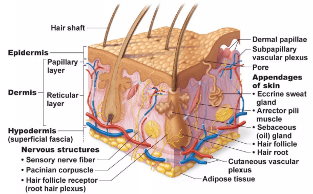
Layers of the skin:
- Epidermis: The outermost layer, composed mainly of dead cells, acts as a waterproof barrier to protect the body and does not contain blood vessels.
- Houses melanocytes, which produce and distribute melanin (the pigment responsible for skin color).
- Dermis: Situated beneath the epidermis, it is made of dense connective tissue, allowing the skin to stretch and flex. It contains blood vessels, glands, and hair follicles.
- Hypodermis (subcutaneous layer): The layer of fat beneath the dermis, stores fat for energy, and acts as a cushion for the body.
Functions of the hypodermis:
- Protects the body from injury.
- Shields against the invasion of harmful particles.
- Helps conserve water and nutrients.
- Aids in regulating body temperature.
- The skin synthesizes vitamin D when exposed to sunlight.
- Contains nerve endings that detect temperature, pressure, and physical pain.
- Excretes water and minerals such as sodium, chloride, and magnesium through sweat glands.
Skin glands:
- Eccrine glands: Help regulate body temperature and are mostly found on the palms and soles of the feet.
- Release sweat, which is a mix of salt and water, helping maintain the body’s salt and water balance.
- Sweat also contains trace amounts of substances like alcohol, lactic acid, and urea that the body needs to expel.
- Apocrine glands: Primarily located in the armpits and groin, secrete an oily substance containing pheromones.
- These glands release sweat in response to stress, anxiety, fear, or pain.
Homeostasis
- Homeostasis: The body’s ability to maintain internal balance, including regulating body temperature.
- When the body gets too hot:
- Sweat glands secrete sweat, and its evaporation from the skin creates a cooling effect.
- Blood vessels in the skin dilate, bringing blood closer to the surface to release heat before it circulates back deeper into the body.
- When the body gets too cold:
- Blood vessels constrict to minimize blood flow to the skin’s surface, conserving heat.
Pathologies
- Acne: A condition where hair follicles become blocked with oil and dead skin cells, leading to inflammation.
- Carcinoma: Skin cancer that originates in the skin’s epithelial tissue, the most common type of skin cancer.
The Urinary System
- Responsible for removing water and waste from the body while maintaining electrolyte balance.
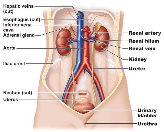
Key components and their roles:
- Kidneys: The primary organs of the urinary system.
- Filter waste and toxins from the blood.
- Maintain electrolyte balance, regulate blood volume, blood pressure, and pH levels.
- Secrete hormones:
- Renin: Helps manage blood pressure by controlling water and salt retention.
- Calcitriol: The active form of vitamin D.
- Regions of the kidney:
- Renal cortex: The outermost layer; contains blood vessels and produces erythropoietin (stimulates red blood cell production).
- Renal medulla: The innermost layer; controls the concentration of urine.
- Renal artery: Delivers oxygen-rich blood to the kidneys.
- Renal vein: Transports filtered, deoxygenated blood away from the kidneys.
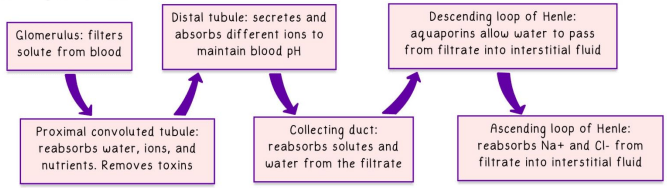
- Nephron: The kidney’s functional unit, consisting of looping tubes that filter electrolytes, metabolic waste, and water-soluble molecules from the blood.
- Urea: A nitrogen-based waste product from protein breakdown.
- Uric acid: A waste product from nucleic acid metabolism.
- Filtrate: The fluid filtered from the blood, containing water, urea, glucose, salts, and other molecules.
- Ureters: Thin tubes that transport urine from the kidneys to the bladder.
- Urethra: Transfers urine from the bladder to outside the body.
- The female urethra is shorter than the male urethra, increasing susceptibility to urinary tract infections.
- Urine: A mix of waste products expelled from the body:
- Bladder: Holds 400–800 ml of urine.
- For urine to exit, two sphincters must open:
- Internal sphincter: Made of smooth, involuntary muscle.
- External sphincter: Controlled voluntarily.
Pathologies
- Urinary tract infection (UTI): An infection in the kidney, bladder, or urethra, more prevalent in women.
- Kidney stones: Hard deposits formed in the kidneys, often painful to pass, usually composed of minerals and acid salts.
