Dermatology Assessment Guidelines
- Evaluate the whole patient, not just the skin condition. Assess for possible transmission or contagion risk.
- Key questions to ask:
- Where did the issue first appear? (Face, torso, extremities, genitals)
- How long has it been present?
- Does it itch?
- Is the patient otherwise healthy?
- Conditions like rosacea, keratosis pilaris, and seborrheic dermatitis are often skin-limited.
- Is the patient uncomfortable but not systemically ill?
- Itching, burning, pain → Could indicate scabies or shingles.
- Is the patient systemically unwell (fever, fatigue, appetite loss, weight loss, malaise)?
- Possible conditions: Varicella, toxic epidermal necrolysis, Lyme disease, lupus.
- Are there primary lesions only, or primary and secondary?
- Where did the oldest lesion first appear, and when?
- Where did the newest lesion appear, and when?
- Primary lesions: Direct result of disease, not altered by external factors.
- Example: Vesicle (<1 cm, fluid-filled) – seen in varicella, herpes, shingles.
- Secondary lesions: Altered by treatment or disease progression.
- Example: Crust – formed by dried serum/blood when a vesicle ruptures.
Smallpox
- Declared eradicated in 1977.
- Infects respiratory and oropharyngeal surfaces; 2-week incubation period.
- Symptoms mimic flu, with large nodules primarily on the face, arms, and legs.
- Mortality rate: 20-50%.
- Post-exposure vaccine (within 3-4 days) can reduce severity.
Common Skin Lesions & Their Characteristics
| Lesion Type | Description | Example Conditions |
|---|---|---|
| Annular | Ring-shaped with central clearing | Lyme disease (bull’s eye lesion) |
| Bulla | Fluid-filled blister >1 cm | Burns |
| Clustered | Grouped lesions without a specific pattern | Herpes |
| Confluent/Coalescent | Merging lesions forming larger patches | Psoriasis |
| Cyst | Encapsulated, raised, fluid-filled lesion | Intradermal cyst |
| Lichenification | Thickened skin from chronic itching/rubbing | Eczema |
| Linear | Appears in streaks | Poison ivy |
| Macule | Flat, non-palpable discoloration <1 cm | Freckle |
| Maculopapular | Flat discoloration with small raised papules | Viral exanthem |
| Nodule | Solid, raised lesion 0.5–2 cm (>2 cm = tumor) | Lipoma |
| Papule | Small, solid elevation <0.5 cm | Mole |
| Patch | Flat discoloration >1 cm | Vitiligo |
| Petechiae | Small red-purple spots <1 cm, do not blanch | Thrombocytopenia |
| Plaque | Elevated, flat lesion >1 cm | Psoriasis |
| Purpura | Red-purple discoloration that doesn’t blanch | Meningococcemia |
| Pustule | Small vesicle filled with pus | Impetigo |
| Reticular | Net-like pattern | Livedo reticularis |
| Scale | Flaky, superficial lesion | Dandruff, psoriasis |
| Scattered | Randomly distributed lesions | Rubella, roseola |
| Vesicle | Clear fluid-filled lesion <1 cm | Herpes |
| Wheal | Swollen, red, raised lesion from edema | Urticaria (hives) |
Skin Cancer Assessment – ABCDE Rule
- A – Asymmetry
- B – Border irregularity
- C – Color variation (brown, black, red, white, blue)
- D – Diameter >6mm (pencil eraser size)
- E – Evolving (changing shape, color, or elevation)
- Melanoma: Dark, uneven-textured moles; may itch.
- Acral Lentiginous Melanoma: More common in African Americans and Asians; often appears on palms, soles, or nail beds.
- Subungual Hematoma: Nailbed trauma causes bleeding under nail; may require trephination (drainage).
Pressure Ulcer Staging
| Stage | Description |
|---|---|
| Stage 1 | Non-blanchable redness on intact skin |
| Stage 2 | Partial-thickness skin loss; may appear as an intact blister |
| Stage 3 | Full-thickness skin loss, exposing fat; crater-like appearance |
| Stage 4 | Full-thickness tissue loss, exposing muscle, bone, or tendons |
Overview of Common Skin Rashes
| Condition | Key Features |
|---|---|
| Impetigo | Golden-yellow crusts on fragile blisters. Intensely itchy. |
| Measles | Koplik’s spots—tiny white lesions on a reddish base, found inside the cheeks near the molars. |
| Scabies | Severe nighttime itching. Wavy, thread-like rash found between fingers, around the waist, underarms, and genital area. |
| Scarlet Fever | Rough, sandpaper-like rash with an accompanying sore throat, commonly caused by Streptococcus bacteria. |
| Tinea Versicolor | Light-colored, round-to-oval patches mainly on the upper back and shoulders. Typically non-itchy. |
| Pityriasis Rosea | Rash follows natural skin folds, forming a “Christmas tree” pattern. Starts with a single, larger “herald patch.” |
| Molluscum Contagiosum | Smooth, dome-like bumps (~5 mm), each with a central dimple and a waxy core. |
| Erythema Migrans | Expanding, bullseye-shaped red rash with central clearing—an early indicator of Lyme disease. |
| Meningococcemia | Painful, dark red or purplish skin spots spreading rapidly. Sudden fever, headache, and confusion. Close contacts may need Rifampin prophylaxis. |
Topical Steroid Potency & Application
- Ranked from Class 7 (least potent) to Class 1 (most potent)
- Application by Body Area:
- Non-folded areas (arms, legs, torso): Triamcinolone 0.1%
- Face & skin folds: Desonide or hydrocortisone
- Palms & soles: Fluocinolone or clobetasol
- Formulation Absorption & Properties:
- Lotions < Creams < Gels < Ointments (in increasing order of potency)
- Creams: Well-absorbed, non-greasy; may sting on broken skin
- Ointments: Rich in emollients; enhances absorption
- Lotions: Water-based, spread easily; mildest option
- Absorption Rates by Area:
- Highest: Face → Next: Underarms & genital region
- General Dosage Guidelines:
- 2g covers hands, face, head, and anogenital area
- 3g covers one arm, front or back of the torso
- 30-60g needed for full-body coverage
Burn Classification & Management
- First-degree burns: Red, blanches with light pressure
- Second-degree burns (partial thickness): Moist, red with peeling edges and scattered blisters (Treat with Silvadene or Polysporin)
- Third-degree burns (full thickness): Thick, pale, or waxy appearance
Referral Criteria:
- Burns on face, hands, feet, genitals, or major joints
- Electrical or lightning-related burns
- Partial-thickness burns covering more than 10% of body surface area (BSA)
- Any full-thickness (third-degree) burn, regardless of age
- Encircling burns (circumferential)
Common Infectious Risks: Pseudomonas aeruginosa, Escherichia coli, Klebsiella pneumoniae
Topical Steroid Potency Chart – High Strength
| Active Ingredient | Potency | Available Forms & Sizes | Brand Names |
|---|---|---|---|
| Betamethasone Dipropionate 0.05% | 100–150× hydrocortisone | Cream/Ointment: 15g, 50g | Diprosone, Beta |
| Lotion: 50mL | Betnovate | ||
| Topical Solution: 100mL | Beta | ||
| Betamethasone Valerate 0.1% | 100–150× hydrocortisone | Cream/Ointment: 15g, 50g | Diprosone, Beta |
| Lotion: 50g | Beta | ||
| Diflucortolone Valerate 0.1% | 100–150× hydrocortisone | Cream/Ointment: 50g | Nerisone |
| Hydrocortisone Butyrate 0.1% | 100–150× hydrocortisone | Lotion: 100mL | Locoid Scalp |
| Emulsion: 100mL | Locoid Crelo | ||
| Cream/Ointment: 30g, 100g | Locoid Lipocream, Locoid Ointment | ||
| Methylprednisolone Aceponate 0.1% | 100–150× hydrocortisone | Not specified | |
| Mometasone Furoate 0.1% | 100–150× hydrocortisone | Lotion: 30mL | Elocon |
| Cream/Ointment: 15g, 50g | Elocon, Advantan, Advaritan |

Skin Conditions
- Fifth Disease – Lace-patterned maculopapular rash
- Varicella, Zoster, Herpes Simplex – Rash with papules, vesicles, and crusts on a red base
- Pityriasis Rosea – Oval, maculopapular lesions with a “herald patch” forming a Christmas tree pattern
- Seborrheic Keratosis – Soft, wart-like lesions on the trunk, varying in color from light brown to black, with a “stuck-on” appearance
- Xanthelasma – Yellowish, raised plaques near the eyelids or brows, often linked to high cholesterol
- Melasma – “Pregnancy mask” appearing as darkened patches on the forehead and upper cheeks
- Vitiligo – Irregular hypopigmented patches on the skin
- Cherry Angioma – Small, bright red papules that blanch under pressure, non-malignant
- Lipoma – Soft, fatty tumors beneath the skin, typically on the neck, trunk, arms, or legs
- Nevi (Moles) – Round, pigmented spots or raised lesions, generally harmless
- Xerosis – Genetically inherited extreme dryness affecting skin, eyes, and mouth
- Acanthosis Nigricans – Velvety, darkened skin thickening, commonly on the neck or armpits, associated with diabetes, obesity, metabolic syndrome, and some cancers
- Acrochordon (Skin Tags) – Soft, flesh-colored, painless skin growths with a pedunculated shape
- Candida Infection – Bright-red rash with satellite lesions, commonly found in moist areas
- Intertrigo – Widespread red rash in skin folds due to bacterial infection
- Acral Lesions – Rashes occurring on extremities
Wound Healing & Treatment
Factors That Slow Healing
- Aging, malnutrition, immune system compromise, limited mobility, chronic stress, diabetes, medications (steroids, anticoagulants), prolonged pressure, smoking, secondary infections
Wound Closure Types
- Primary Healing – Closed within 24 hours using sutures, adhesive strips, or tissue glue
- Secondary Healing – Left open to heal gradually from the inside out
- Tertiary Healing – Heavily contaminated or poorly perfused wounds (e.g., crush injuries) left open initially before closure
Referral Criteria
- Infected wounds, clenched-fist injuries, facial wounds with potential cosmetic concerns, retained foreign objects, joint capsule involvement, electrical or chemical burns, high-pressure injection wounds, suspected child abuse
Wound Care Guidelines
- Avoid suturing wounds older than 24 hours (infection risk increases after 12 hours)
- Tdap booster required if last tetanus shot was over 5 years ago
- Suture Removal Timeline
- Face: 5–7 days
- Scalp: 7–10 days
- Upper limbs: 7–10 days
- Lower limbs: 10–14 days
Miscellaneous Medical Notes
- Bed Bugs – Cause irritation but do not spread infections
- Squamous Cell Carcinoma (SCC)
- Common in sun-exposed areas, lower lip frequent in smokers
- Appears as papules, plaques, nodules, smooth or ulcerated lesions, often bleeds easily
- Diagnosis confirmed via biopsy or excision
- “NOSUN” Mnemonic for SCC Features:
- N – Nodular
- O – Opaque
- S – Sun-exposed areas
- U – Ulcerative
- N – Non-distinct borders
- Irritant Contact Dermatitis – Triggered by prolonged exposure to water, soaps, detergents, fiberglass, dust, food, cleaning agents, lubricants, petroleum products, solvents, and resins
- Allergic Contact Dermatitis – Caused by allergens such as poison ivy, rubber, nickel, and fragrances
Lichen Planus
- Appearance: Small, flat, reddish-purple bumps with white scaling
- Symptoms: Itching, commonly affecting wrists, forearms, and ankles
- Causes: Associated with Hepatitis C; usually self-resolves
Antibiotic Guide for MRSA (ABCD Mnemonic)
- A – Antibiotics for MRSA
- B – Bactrim
- C – Clindamycin
- D – Doxycycline
| Condition | Cause | Symptoms | Diagnosis | Treatment | Key Considerations |
|---|---|---|---|---|---|
| Rocky Mountain Spotted Fever | Bite from infected dog or wood tick (Rickettsia rickettsii) | Sudden high fever, chills, severe headache, nausea, vomiting, light sensitivity, muscle pain, joint pain, conjunctivitis. Rash (small red spots) appears 2-5 days later, starting on hands/feet and spreading to the trunk. | Antibody testing for Rickettsia, skin biopsy, bloodwork (CBC, LFTs, CSF analysis). | Doxycycline 100 mg twice daily for 7-14 days. Early treatment is critical—can be fatal if delayed past 8 days. Remove tick carefully by gripping near the skin and pulling steadily upward. | Fatal in 3-9% of cases. Most common in southeastern and south-central U.S. (April-September). |
| Brown Recluse Spider Bite | Fever, chills, nausea, vomiting. “Red, white, and blue” lesion: central blister with surrounding gray/purple discoloration, encircled by pale skin and redness. | Apply ice immediately. Clean wound, apply antibiotic ointment, elevate affected area, limit movement, take NSAIDs for pain. Prevention: check shoes, boxes, and storage areas before reaching inside. | Found in the Midwest and Southeast U.S. Severe cases may lead to necrosis. | ||
| Erythema Migrans (Lyme Disease) | Infection with Borrelia burgdorferi from a deer tick bite | Expanding red rash with a central clearing (“bullseye”), warm to the touch with a rough texture. Flu-like symptoms. Rash appears 7-14 days after bite and resolves on its own. | Enzyme immunoassay (EIA), indirect immunofluorescence assay (IFA). | Early treatment: Doxycycline twice daily for 14-21 days (up to 28 days). Alternatives: Amoxicillin or Ceftin. Avoid doxycycline in children (stains teeth). | Can cause systemic infection, heart block, Bell’s palsy, Guillain-Barré, and chronic joint pain. Common in Northeast U.S. |
| Meningococcemia | Neisseria meningitidis (Gram-negative bacteria, respiratory transmission) | Sudden sore throat, cough, fever, headache, stiff neck, light sensitivity, altered mental state. Rapid development of petechial or hemorrhagic rash. | Lumbar puncture (CSF analysis), blood/throat cultures, brain imaging (CT/MRI). | Immediate hospitalization. IV Rocephin (2g every 12h) + IV Vancomycin (every 12h). Isolation and supportive care. Preventive treatment for close contacts: Rifampin twice daily for 2 days and meningococcal vaccine. | Life-threatening within 48 hours. High risk for college dorm residents, people with asplenia, sickle cell disease, or HIV. |
| Varicella / Zoster | Varicella-zoster virus (chickenpox/shingles, spread by inhalation or direct contact) | Chickenpox: Fever, sore throat, fatigue, itchy blisters that start on the head and spread to the trunk (various stages: vesicles, pustules, crusts). Shingles: Painful grouped vesicular rash along a nerve pathway (dermatome). Contagious until lesions crust over. | Viral culture, polymerase chain reaction (PCR) test, Tzanck smear for shingles. | Chickenpox: Acyclovir within 24-48 hours (avoid aspirin & NSAIDs). Shingles: Acyclovir or Valacyclovir for 7-10 days. | Shingles can lead to long-term nerve pain (postherpetic neuralgia). Risk of eye involvement (herpes zoster ophthalmicus) causing vision loss. |
| Malignant Melanoma | UV exposure, genetic predisposition | Asymmetry, Border irregularity, Color variation (brown, black, red, white, blue), Diameter >6mm, Evolving shape/size. May be itchy. | Skin biopsy (gold standard). | Immediate dermatology referral. Treatment depends on stage: surgical removal, chemotherapy, or immunotherapy. | High risk of spreading (metastasis). More common in fair-skinned individuals, those with frequent sun exposure, or a family history. |
| Basal Cell Carcinoma | UV exposure, fair skin | Waxy, pearly nodule with well-defined edges. May have small visible blood vessels (telangiectasia). Slow-growing, may ulcerate. “PUT ON” mnemonic: Pearly, Ulcerating, Telangiectasia, On sun-exposed areas, Nodule. | Skin biopsy. | Surgical excision, Mohs micrographic surgery. | Most common skin cancer. Low risk of spreading but can cause severe local tissue damage if untreated. |
| Actinic Keratosis | Small, rough, scaly patches on sun-exposed areas (face, ears, scalp, hands). Can range from tiny spots to large lesions. | Clinical diagnosis. | 5-Fluorouracil (5FU) cream, imiquimod, topical diclofenac gel, liquid nitrogen, laser therapy, or chemical peels. Dermatology referral for biopsy if suspicious. | Considered pre-cancerous. Can develop into squamous cell carcinoma. | |
| Erythema Multiforme (Stevens-Johnson Syndrome) | Severe allergic reaction (NSAIDs, sulfa drugs, anti-epileptics), infections (Herpes simplex, Mycoplasma pneumoniae), malignancies | Sudden eruption of red “target” lesions with blistering and peeling skin. Accompanied by fever, flu-like symptoms 1-3 days before rash appears. | Risk of progression to Toxic Epidermal Necrolysis (TEN), a life-threatening condition if >30% of the skin is affected. |
| Condition | Cause | Signs & Symptoms | Diagnostic Methods | Treatment Options | Key Considerations |
|---|---|---|---|---|---|
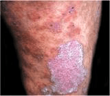
| Psoriasis | Genetic predisposition; rapid skin cell turnover | Red, scaly plaques with fine silvery scales, typically on elbows, scalp, knees, and gluteal folds. Nail pitting. Auspitz sign: pinpoint bleeding when scales are removed. Koebner phenomenon: new lesions develop at sites of skin trauma. | Clinical diagnosis | First-line: Medium-strength topical corticosteroids. Second-line: Vitamin D analogs, topical retinoids (tazarotene), coal tar. UVB therapy may help. Caution: Beta-blockers can worsen symptoms. | Can lead to psoriatic arthritis (joint pain, swelling, warmth) and guttate psoriasis (linked to Group A strep infection). |
| Tinea Versicolor | Fungal infection by Pityrosporum orbiculare or Pityrosporum ovale | Hypopigmented, round macules on the chest, shoulders, and back that become noticeable after tanning. Usually asymptomatic. | KOH prep: Shows “spaghetti & meatballs” pattern (hyphae & spores). | Topical: Selenium sulfide, ketoconazole cream. Oral: Antifungal medications if severe. | Common in warm, humid climates. Can recur despite treatment. |
| Atopic Dermatitis (Eczema) | Genetic condition causing chronic itchy rash | Location: Hands, flexural folds, neck. Appearance: Itchy, red, round-to-oval plaques that may ooze and later become thickened (lichenified). Exacerbated by stress and environmental factors. | Clinical evaluation | Moisturizers, lukewarm baths, and topical corticosteroids (low to high potency). Oral antihistamines for itching. | Risk of skin fissures and infections. Often part of the “atopic triad” (eczema, allergies, asthma). |
| Acute Cellulitis | Bacterial infection (Strep pyogenes, Staph aureus including MRSA) | Red, swollen, warm, and tender skin infection with poorly defined edges. | Clinical assessment | Non-purulent: Cephalexin or dicloxacillin. MRSA suspected: Bactrim, doxycycline, or clindamycin. Tetanus booster if >5 years since last dose. | Monitor closely—follow up in 48 hours. Refer if worsening, spreading, or affecting immunocompromised patients. Possible complications: osteomyelitis, sepsis. |
| Cutaneous Abscess, Furuncle, Carbuncle | Staph aureus (MSSA or MRSA) | Infection of a hair follicle leading to a painful, red lump (abscess). Carbuncle: Cluster of abscesses. | Culture & Sensitivity (C&S) | Incision & drainage (I&D), warm compresses. Antibiotics: Bactrim, doxycycline, or clindamycin. MSSA: Dicloxacillin or cephalexin. | Consider mupirocin (Bactroban) for folliculitis to prevent recurrence. |
| Erysipelas | Strep pyogenes | Rapid-onset red, swollen, warm skin with sharp borders, commonly on legs or face. | Clinical diagnosis | Mild cases: Keflex or dicloxacillin. Severe cases or immunocompromised patients: Hospitalization for IV antibiotics. | Can spread rapidly. Immediate treatment reduces risk of complications. |
| Bite Wounds | Animals: Pasteurella multocida (dogs/cats). Humans: Eikenella corrodens. Rabies risk: Skunks, raccoons, foxes, coyotes. | Human bites: Highest infection risk. Cat bites more likely to cause infection than dog bites. Rabies risk with wild animal bites. | Wound culture & sensitivity | First-line: Augmentin for 10 days. PCN allergy: Doxycycline or Bactrim + Flagyl/clindamycin. Rabies protocol: Immune globulin + vaccine if indicated. | Do not suture infected wounds. Follow-up in 24-48 hours. 80% of cat bites become infected. |
| Hidradenitis Suppurativa | Chronic inflammation of sweat glands (often in axilla, groin) | Painful, deep red nodules under the arms or in the groin that may rupture and form abscesses. | Culture & Sensitivity of drainage | Mild: Chlorhexidine washes, topical clindamycin for 12 weeks. Moderate-severe: Oral tetracyclines, doxycycline, or minocycline. | High recurrence rate. Can lead to scarring and sinus tract formation. |
| Impetigo | Staph aureus or Strep pyogenes | Non-bullous: Red sores that develop into honey-colored crusts. Bullous: Blisters filled with clear or yellow fluid that rupture, leaving raw skin. Ecthyma: Deep ulcerated form. | Culture & Sensitivity of crusts or lesions | Mild cases: Mupirocin 2% ointment for 10 days. Widespread infection: Cephalexin or dicloxacillin. PCN allergy: Azithromycin or clindamycin. | Highly contagious. No school for 48-72 hours after starting treatment. More common in warm, humid weather. |
| Condition | Cause | Signs & Symptoms | Diagnostics | Treatment | Key Considerations |
|---|---|---|---|---|---|
| Herpetic Whitlow | Herpes simplex virus (HSV-1 or HSV-2) | Painful blisters on the side of the finger or near the cuticle. Often follows direct contact with a cold sore or genital herpes lesion. | Clinical diagnosis | Pain relief with NSAIDs or analgesics. Severe cases: Acyclovir. | Avoid sharing personal items (gloves, towels). Keep lesions covered until fully healed to prevent spread. |
| Pityriasis Rosea | Unknown (suspected viral origin) | “Herald patch”: single oval lesion with a salmon-colored center and red outer ring on the trunk. Later spreads in a “Christmas tree” pattern along skin lines. | Clinical diagnosis; rule out secondary syphilis if needed. | Self-limiting (6-8 weeks). If itchy: Antihistamines or mild topical steroids. | Benign condition; no treatment needed unless symptomatic. |
| Scabies | Sarcoptes scabiei (mite infestation) | Intensely itchy rash, worse at night. Common in web spaces of fingers, toes, axillae, waist, groin, buttocks, breasts, and penis. Linear burrows may be visible. | Skin scraping + wet mount: Identifies mites, eggs, or feces under a microscope. | First-line: Permethrin 5% cream (apply to entire body, wash off after 8-12 hours). Household contacts must be treated. Wash clothes and bedding in hot water. | Avoid Lindane (Kwell) due to neurotoxicity risk. Symptoms may persist for weeks after treatment. |
| Tinea (Ringworm) / Dermatophytosis | Fungal infection | Types: Scalp (tinea capitis), feet (tinea pedis), body (tinea corporis), groin (tinea cruris), hands (tinea manuum), beard (tinea barbae). Presentation: Itchy, red, circular patches with central clearing and raised, scaly edges. | KOH prep: Reveals fungal hyphae and spores. | First-line: OTC antifungal creams (azole-based or terbinafine). Severe cases: Oral antifungals (e.g., griseofulvin—monitor liver function). | Tinea capitis requires oral treatment. Can recur if hygiene measures aren’t followed. |
| Onychomycosis (Fungal Nail Infection) | Fungal infection (yeast or dermatophytes) | Thickened, yellow, brittle nails. Most commonly affects the great toenail. Onycholysis: Nail separates from the nail bed. | Nail culture or KOH prep | Oral antifungals: Fluconazole (weekly for 2-3 months) or terbinafine (Lamisil). Monitor liver enzymes (LFTs) before and during treatment. | Long treatment duration (months). High recurrence rate. |
| Acne Vulgaris | Hormonal imbalance, excess oil production, bacterial overgrowth, genetics | Lesion types: Open (blackheads), closed (whiteheads), papules, pustules. Severity: – Mild: < 20 comedones, < 15 inflammatory lesions, < 30 total. – Moderate: 20-100 comedones, 15-50 inflammatory, 30-125 total. – Severe: > 5 cysts, extensive involvement. | Clinical evaluation | Mild: OTC washes (salicylic acid, benzoyl peroxide). Moderate: Topical retinoids, antibiotics (clindamycin, tetracycline). Severe: Oral isotretinoin (Accutane), steroid injections. For hormonal cases: Oral contraceptives (Yaz, Desogen). | Takes 6-8 weeks for noticeable improvement. Tetracyclines stain teeth in children under 13. Accutane is teratogenic—requires two forms of birth control. |
| Rosacea | Chronic inflammatory skin condition | Redness, flushing, small papules and pustules around the nose, mouth, and chin. Ocular symptoms (dry eyes). More common in individuals with fair skin and light eyes (Celtic background). | Clinical diagnosis | First-line: Lifestyle modifications (avoid triggers: spicy food, alcohol, sunlight). Meds: Metronidazole gel (Metrogel), azelaic acid, or low-dose tetracyclines. | Complications: – Rhinophyma: Thickening of nose tissue. – Ocular rosacea: Involves eyelids and conjunctiva (blepharitis, redness). |
| Anthrax | Bacillus anthracis (bacterial infection; zoonotic or bioterrorism exposure) | Cutaneous form: Papule enlarges within 24-48 hours, forming a black eschar with surrounding redness and swelling. Pulmonary form: Flu-like symptoms progressing to severe cough, chest pain, hemoptysis, dyspnea, hypoxia, and shock. | Exposure history + bacterial culture. Pulmonary form: Chest X-ray or CT scan may show mediastinal widening. | Cutaneous form: Doxycycline, ciprofloxacin, or levofloxacin (7-10 days). Bioterrorism exposure: Ciprofloxacin 500 mg BID for 60 days. | High mortality rate for pulmonary anthrax. Immediate medical intervention required for suspected exposure. |
